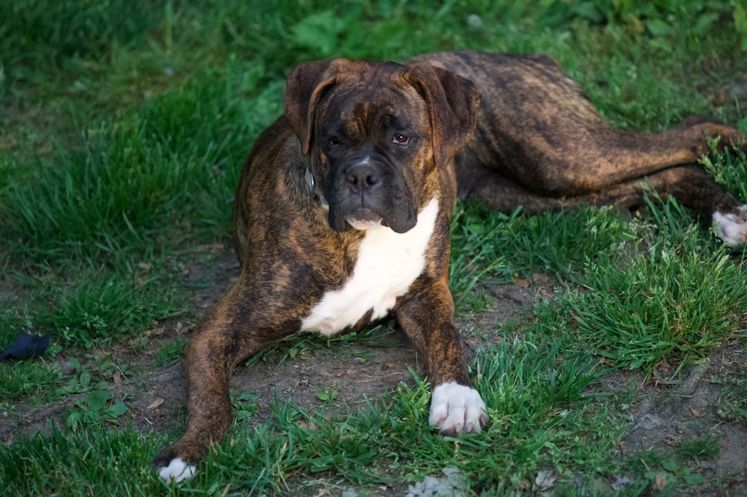
When your large-breed dog starts dragging a foot or struggling to rise, the instinct is to assume degenerative myelopathy — but lumbosacral disease can look almost identical, and unlike DM, it’s a condition your vet may actually be able to treat.
What Is Lumbosacral Disease in Dogs?
Lumbosacral disease is a compression problem at the very base of the spine, where the last lumbar vertebra meets the sacrum. That junction is home to a bundle of nerve roots called the cauda equina — Latin for “horse’s tail” — which controls the hind limbs, bladder, bowel, and tail. When that junction degenerates, narrows, or becomes unstable, those nerve roots get compressed, and the dog starts showing the same kinds of hind-end symptoms you’d associate with DM or IVDD.
The underlying causes can include intervertebral disc degeneration at L7-S1, bony overgrowth (spondylosis), instability at the lumbosacral junction, or a combination of all three. The result is nerve compression that produces weakness, dragging, tail dysfunction, and sometimes severe pain.
German Shepherds are among the breeds most commonly diagnosed, which is particularly significant because German Shepherds are also one of the breeds most commonly affected by DM. That overlap creates a real diagnostic trap for owners and sometimes even for vets who don’t have advanced imaging available.
- The cauda equina is the bundle of nerve roots at the end of the spinal cord, running through the lumbosacral junction
- These nerves control the hind legs, bladder, bowel, and tail
- Compression of this bundle — from disc disease, bony changes, or instability — is what causes cauda equina syndrome
- The condition goes by several names: lumbosacral disease, lumbosacral stenosis, cauda equina syndrome, and degenerative lumbosacral stenosis (DLSS)
How Do DM and Lumbosacral Disease Overlap?
Both conditions appear most often in middle-aged to older large-breed dogs. Both can cause dragging of the hind feet, weakness when rising, stumbling, and changes to how the dog holds or uses its tail. If you looked at a video of each dog without any other context, you might not be able to tell them apart.
The overlapping signs include:
- Hind limb weakness: Both conditions affect the nerves controlling the back legs, resulting in knuckling, stumbling, or a wide-based stance
- Dragging: Paw scuffing and toe dragging occur in both — the underlying mechanism differs, but the visible result is similar
- Tail changes: Reduced tail wagging, a low-hanging or limp tail, or loss of tail tone can appear in either condition
- Difficulty rising: Both dogs may struggle to get up from a lying position, especially on slick surfaces
- Bladder and bowel changes: In advanced stages, both can affect urinary and fecal continence
This symptom overlap is exactly why lumbosacral disease is one of the most common misdiagnoses when owners and vets are considering DM — and it’s an issue the DM vs IVDD comparison and DM vs Wobbler Syndrome articles on this site touch on from different angles.
What Are the Key Differences Between DM and Lumbosacral Disease?
The clearest clinical differentiators are pain, response to treatment, and what imaging reveals. DM and lumbosacral disease diverge sharply on all three.
Pain: The Most Important Clue
Lumbosacral disease is frequently painful. Dogs with cauda equina compression often show pain when you press on the lower back or base of the tail, may cry out when sitting down or jumping, resist being touched in that region, and sometimes show a tucked posture or difficulty finding a comfortable position.
DM, in contrast, is widely considered a painless condition. A dog with pure DM should not flinch when the lumbosacral area is palpated. If your dog is clearly in pain, lumbosacral disease or another structural cause deserves serious consideration — even if DM is also a possibility given the breed or genetic testing.
Response to Rest and Anti-Inflammatories
This is where the clinical picture diverges most usefully. Dogs with lumbosacral disease often show meaningful improvement with a period of rest and a course of anti-inflammatory medication. The nerve compression is mechanical, and reducing inflammation around the compression point can produce real, observable relief — sometimes within days to a few weeks.
DM does not respond to anti-inflammatories, rest, or any currently available medication. A dog whose weakness visibly improves after a week of NSAIDs and restricted activity is almost certainly not dealing with pure DM. That treatment response — or lack of it — is one of the most practical clues available before imaging is in hand.
Imaging Findings
MRI is the gold standard for diagnosing lumbosacral disease, though CT also provides useful information about bony changes. Imaging in cauda equina syndrome typically reveals narrowing at the L7-S1 junction, disc material compressing the nerve roots, or instability and malalignment at that joint. These are structural, visible findings.
DM does not produce visible abnormalities on MRI or CT in the spinal cord itself — the neurodegeneration happens at a cellular level that imaging cannot detect. A DM diagnosis is, in part, a diagnosis of exclusion: imaging is used to rule out compressive or structural causes, not to confirm DM directly. The Merck Veterinary Manual describes DM as a diagnosis that requires ruling out other conditions with similar presentations, which is exactly why imaging matters so much in these cases.
- Pain when touching the lower back or tail base
- Crying out when sitting, lying down, or being lifted
- Visible improvement with rest and anti-inflammatory medication
- Sudden onset of symptoms rather than gradual progression over months
- Asymmetric weakness (one side noticeably worse than the other early on)
Why Getting the Diagnosis Right Matters So Much
This is not just an academic distinction. The two conditions have completely different care paths.
Lumbosacral disease has real treatment options. Conservative management — controlled rest, weight management, physical rehabilitation, and anti-inflammatory medications — helps many dogs with mild to moderate cauda equina compression. When conservative care isn’t enough, surgical decompression at the lumbosacral junction is a well-established procedure that many dogs respond to well. The American College of Veterinary Surgeons describes lumbosacral decompression surgery as a viable option for dogs with significant or worsening neurological deficits from cauda equina compression.
DM has no disease-modifying treatment. The focus for a DM dog is quality-of-life preservation: aggressive physical therapy and exercise to slow functional decline, mobility aids, and eventually wheelchair support. Pursuing surgery for a dog with DM rather than lumbosacral disease would be expensive, stressful, and futile. Going the other direction — assuming DM and pursuing only supportive care for a dog that actually had operable lumbosacral disease — could mean missing a real window for meaningful recovery.
From what I’ve heard consistently from caregivers navigating this diagnosis, the dogs who got imaging early fared better, either because lumbosacral disease was found and treated, or because a confirmed DM diagnosis let their families start planning the right support from the beginning. The early signs of DM article covers what the DM progression typically looks like over time, which can help you compare against what you’re actually seeing in your own dog.
- Ask your vet specifically about lumbosacral disease as a differential diagnosis — not just DM
- Request a hands-on neurological exam with palpation of the lumbosacral junction
- If your dog shows any pain signs, push for advanced imaging before assuming DM
- Ask whether a supervised trial of anti-inflammatory medication plus rest is appropriate as a diagnostic tool
- Consider a veterinary neurologist referral if your general vet is uncertain — these cases genuinely benefit from specialist evaluation
What a Definitive Workup Looks Like
A thorough evaluation typically includes a complete neurological exam, assessment for pain at the lumbosacral junction, and ideally MRI or CT imaging. SOD1 genetic testing for the DM mutation can be a useful supporting data point — a dog that tests “at risk” is a better DM candidate, but a negative or “carrier” result should increase suspicion of an alternative diagnosis. The SOD1 genetic test article explains exactly what those results do and don’t tell you.
The honest reality is that some dogs will have both conditions simultaneously — DM affecting the spinal cord while lumbosacral degeneration simultaneously compresses the cauda equina. In those cases, treating the lumbosacral component can still improve quality of life even if the DM continues to progress. No single test closes the door on all possibilities, which is exactly why pushing for a complete workup is worth the effort and the cost.
The caregivers I’ve seen navigate this best are the ones who refused to accept “it’s probably DM, let’s wait and see” when their dog was clearly in pain or when symptoms appeared suddenly rather than over months. Slow, symmetric, painless progression in a high-risk breed is DM’s fingerprint. Pain, asymmetry, and a good response to treatment are lumbosacral disease’s fingerprint. Those aren’t the same dog.
Related Reading
- Early Signs of DM That Owners Often Miss
- Getting a DM Diagnosis: What the Process Actually Looks Like
- The SOD1 Genetic Test for DM: What It Tells You (and What It Doesn’t)
Frequently Asked Questions
Can a dog have both DM and lumbosacral disease at the same time?
Yes, and this is more common than most owners realize — especially in older large breeds. When both conditions are present, it makes diagnosis harder and means your vet needs imaging to understand what’s driving the symptoms. Treatment decisions become more complex, but lumbosacral disease is still often worth addressing even when DM is also confirmed.
How is lumbosacral disease diagnosed in dogs?
Diagnosis typically requires advanced imaging — MRI is the gold standard, though CT is also commonly used. X-rays may hint at changes at the lumbosacral junction but are not definitive. A neurological exam and response to conservative treatment can also provide useful information before imaging is pursued.
Does lumbosacral disease cause pain in dogs the way DM does not?
Yes — pain is one of the clearest differentiators. Lumbosacral disease frequently causes pain at the base of the tail, reluctance to sit or lie down, and resentment when that area is touched. DM is generally considered a painless condition, so a dog showing clear signs of discomfort deserves a closer look for lumbosacral or another structural cause.
What breeds are most at risk for lumbosacral disease?
Large breeds are disproportionately affected, with German Shepherds being one of the most commonly cited — the same breed that’s also at high risk for DM. Other large working breeds including Belgian Malinois, Labrador Retrievers, and Rottweilers are also frequently diagnosed. This breed overlap is a major reason lumbosacral disease and DM are so often confused.
This guide is based on real experience and should be used alongside professional veterinary care. Always consult your veterinarian before starting any new treatment or making changes to your dog’s care plan.